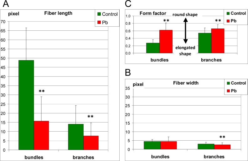
Fig 8. The analysis of morphology of actin filaments Lemna trisulca mesophyll cells.
Determination of the differences in microfilament morphology was carried out based on the measurement of length (A—fiber length), thickness (B—fiber width) and shape (C—form factor) of microfilament bundles and their branches surrounding chloroplast in control plants and plants treated with lead in darkness. In cells treated with lead, disturbances in microfilaments morphology consisted of shortening (A) and rounding (C) both bundles and their branches. The width of microfilaments was lower in plants treated with lead (B) but only in the case of microfilaments branches. **p < 0.05

