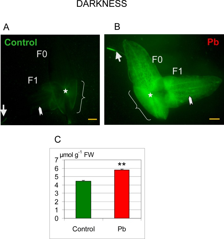
Fig 9. Distribution of hydrogen peroxide in Lemna trisulca.
H2O2 localization by labeling with dichlorodihydrofluorescein diacetate (DCFH-DA; green fluorescence) in fronds (F0—mother fronds and F1—daughter fronds) and in the root of control plants (A) and plants treated with lead (B) in darkness. The control plants showed weak fluorescence in the area of the node (asterisk), sheaths (curly bracket), in the root tip (arrow) and in vascular bundles (arrowhead). Fluorescence intensity was higher in plants treated with lead (B) than in control plants (A). Additionally, under the influence of lead, hydrogen peroxide was generated in mesophyll cells of fronds (double arrow). (C) H2O2 content was also determined spectrometrically in control (Control) plants and plants treated with Pb. **p < 0.05; Scale bar = 1mm

