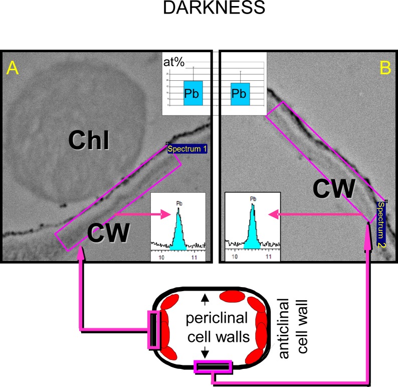
Fig 10. Colocalization of chloroplasts and Pb in Lemna trisulca.
Lack of relationship between the location of chloroplasts and lead deposits in cell walls of plants treated with the metal in darkness. Lead content is shown as atomic percentage (at%, histograms) and as fragments of X-ray spectra (blue color marks the energy range corresponding to lead atoms) based on X-ray map microanalyses performed in areas of the same size (n = 30). Sample areas of such analyses in regions with chloroplasts (A) and without them (B) are shown as pink rectangles on images from transmission electron microscopy. Below, a diagram of a mesophyll cell section is presented, with marked sample areas of the analyses. Chl—chloroplast, CW—cell wall.

