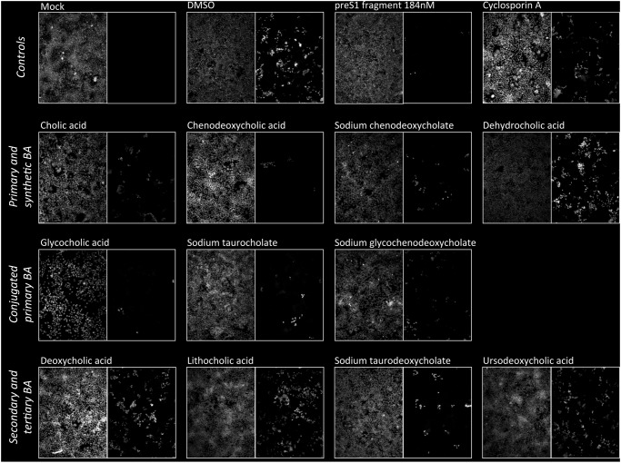
Fig 1. Effect of BA on HDV infection of HuH-7/hNTCP cells.
HuH-7/hNTCP cells were exposed to HDV containing supernatant in the presence or absence of 200 µM of different BA. For glycocholic acid 50 µM results are shown because the BA is toxic at 200 µM. preS1 peptide (368 nM) and cyclosporine A (25 µg/mL) served as positive controls. After 6 h cells were washed and repleted with regular media without virus, BA or drugs. After another 5 d cells were fixed and stained with a polyclonal antibody against HDV antigen (right hand images in each panel). Nuclei were counterstained with DAPI (left hand images). A representative of at least three independent experiments is shown.

