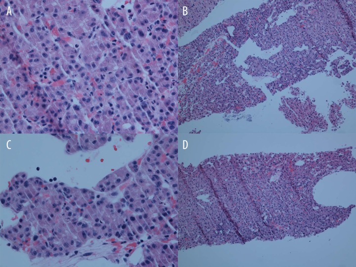
Figure 3.
Histopathologic specimen from the liver biopsy. 10×: A well-differentiated hepatocellular carcinoma with cells in a trabecular pattern and forming pseudo glands. 40×: Malignant hepatocytes with prominent nucleoli and some intracytoplasmic bile. The arrow highlights endothelial wrapping of the tumor cells, which is a feature of HCC.

