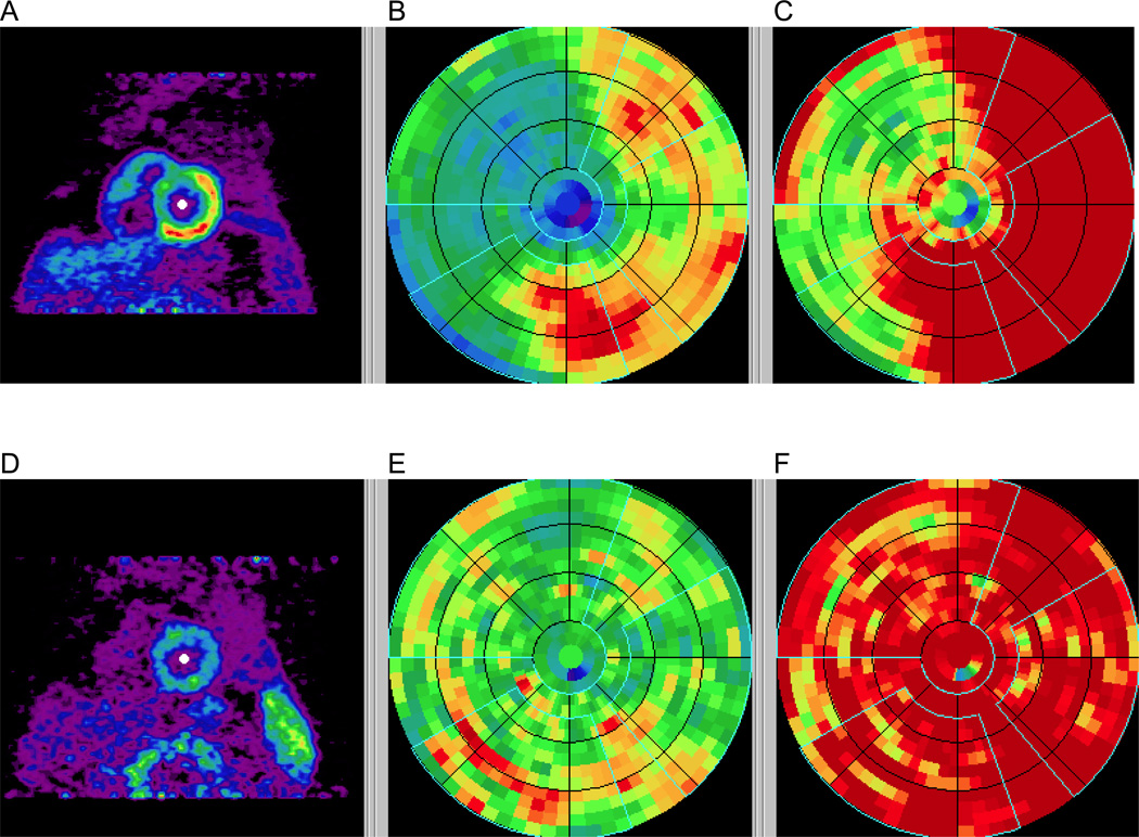
Figure 1.
Polar maps in a CAD patient with stress images (A–C) and rest images (D–F) demonstrating a reversible defect affecting mostly the LAD territory. Early summed images (0.5–2 min) were re-oriented into short-axis views (A, D), polar maps generated (B, E), and normalized based on averages of normal subjects (C, F). The vascular territories and the left ventricular chamber were defined on the polar map automatically.

