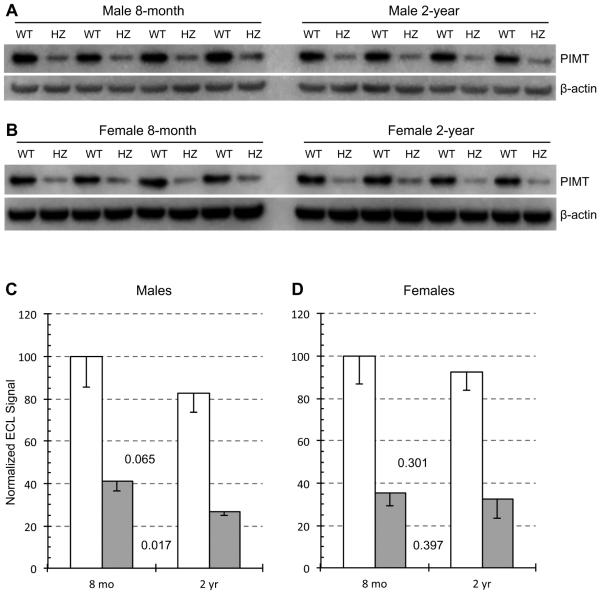
Fig. 3.
Effect of animal age on PIMT expression in brain extracts of WT and HZ mice. Western blots used a mixture of primary antibodies to PIMT and β-actin for male mouse brain extracts (A) and female mouse brain extracts (B). Panels C and D show quantitative measurements of band intensities after normalization to β-actin. Data are expressed as means ± SD (n=4 mice for each group). P-values are based on a two-tailed t-test (paired).

