
Figure 6. SHP-2 activation by fusaruside suppresses p-STAT1/T-bet and production of Th1 cytokines upon TCR stimulation.
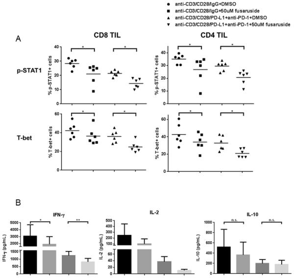
Total TIL were stimulated with anti-CD3/-CD28/hIgG1 beads (bead: cell=10:1) or anti-CD3/-CD28/PD-L1 beads plus 100ug/mL anti-PD-1 blockade (BMS-936558) for 48h in the presence of 50uM fusaruside or DMSO. Then p-STAT1 and T-bet were analyzed by flow cytometry. Supernatants were collected and stored at −80°C. Th1 (IFN-γ and IL-2) and Th2 (IL-10) cytokines in the supernatants were determined by Luminex. A) Summary data of frequency of p-STAT1+ and T-bet+ cells in CD8+ and CD4+ TIL at different conditions is shown (n=6). B) Summary data of amount of IFN-γ (n=8), IL-2 (n=4) and IL-10 (n=8) in the supernatants of TIL cultured under indicated conditions. The graphs present the mean ± SEM from different HNSCC patients. Statistical significance was determined by Wilcoxon (non-parametric paired) test. *p<0.05, **p<0.01. p>0.05 was considered to be not significant (n.s.).