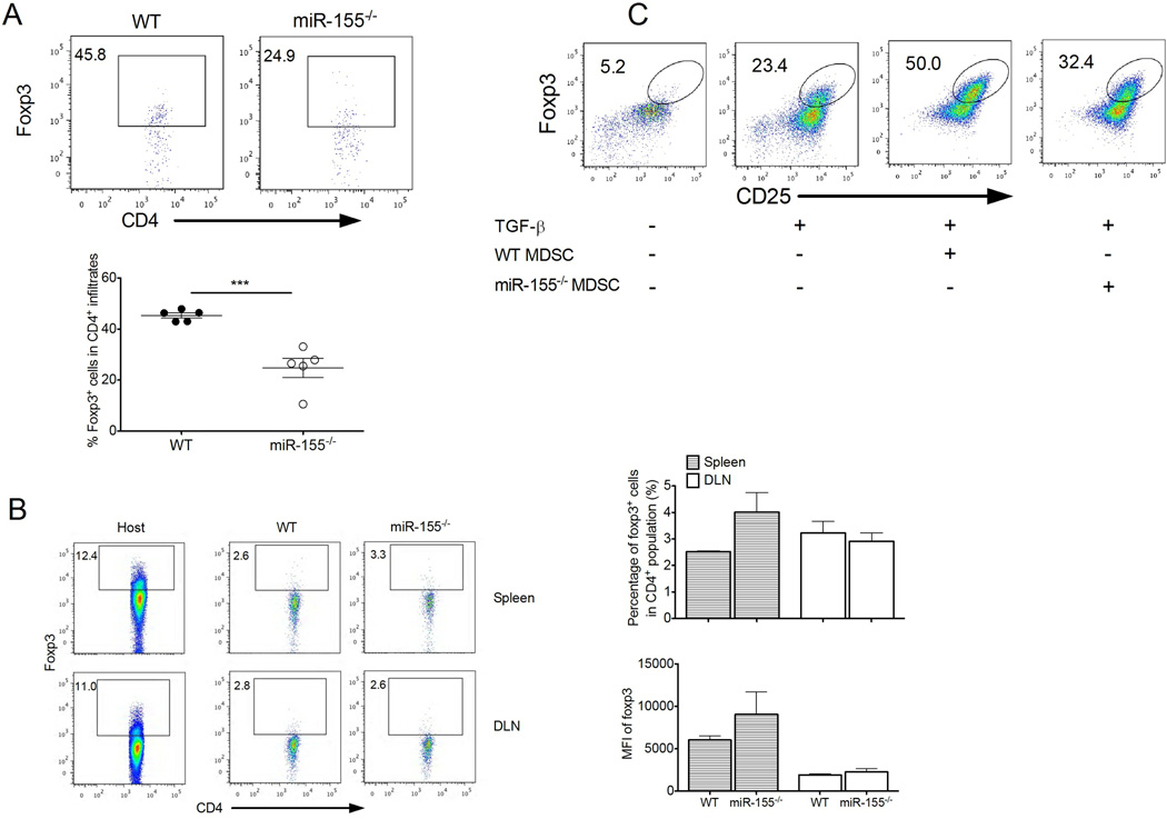
Figure 6. miR-155 is required for MDSC-mediated Treg induction.
(A) Representative dot plots of Foxp3 expression in LLC1-OVA tumor-infiltrating CD4+ cells. Percent Foxp3+ cells is indicated within plots and summarized. (n= 5). (B) WT or miR-155−/− CD4+CD62L+ naïve T cells were transferred into CD45.1 mice followed by a s.c. injection of LLC1-OVA cells. The conversion of transferred T cells to Foxp3+ cells (CD45.2+) in DLN and spleen from LLC1-OVA tumor-bearing mice were detected by flow cytometer 9 d after tumor cell injection. The levels of converted Foxp3 expression are determined by mean fluorescent intensity (MFI). Endogenous Foxp3+ cells (CD45.1+) from host mice were shown as controls. (C) WT and miR-155−/− MDSCs from LLC1 tumor-bearing mice were cocultured with OT-II splenocytes at a 1:4 ratio for 5 days in the absence or presence of TGF-β, and induced CD25+Foxp3+ cells among total CD4+ T cells were subsequently determined by flow cytometry. ***, P<0.001. Data are representative of 2 independent experiments.

