
Figure 1.
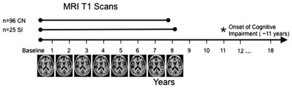
Schematic of study design.
Data were used from the Baltimore Longitudinal Study of Aging, where participants are followed annually for MRI scans, clinical and cognitive testing. The BLSA is currently in it’s 18th year of data collection. From the entire sample of participants, only those who had at least 3 MR evaluations, normal cognitive performance at baseline, and no confounding health conditions were included. These participants were then split into two groups, based on their cognitive diagnosis. 96 participants have remained cognitively normal to date (CN group) and had an average of 7.6 scans per participant included. The other 25 participants have shown subsequent cognitive impairment resulting in a diagnosis of either MCI or AD (SI group). Only scans collected before the onset of cognitive impairment were used for this group, which was an average of 8.0 scans per participant. The subsequent cognitive impairment occurred an average of 11.2 years after the initial scan.