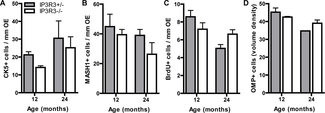
Figure 5. Cell populations and proliferation in aged OE of IP3R3+/− and IP3R3−/− mice.
The numbers of (A) CK5+ horizontal basal cells and (B) MASH1+ globose basal cells in the OE of 12 and 24 month IP3R3+/− and IP3R3−/− mice were comparable. The rates of basal cell proliferation (C) measured by BrdU incorporation were not altered in the OE of IP3R3+/− and IP3R3−/− mice at 12 and 24 months. The volume density of OMP+ neurons (D) was not changed in the OE of IP3R3+/− and IP3R3−/− mice at 12 and 24 months. P > 0.05; Two-way ANOVA followed by Tukey/Kramer Procedure post-hoc test; n = 3–6 mice per group.

