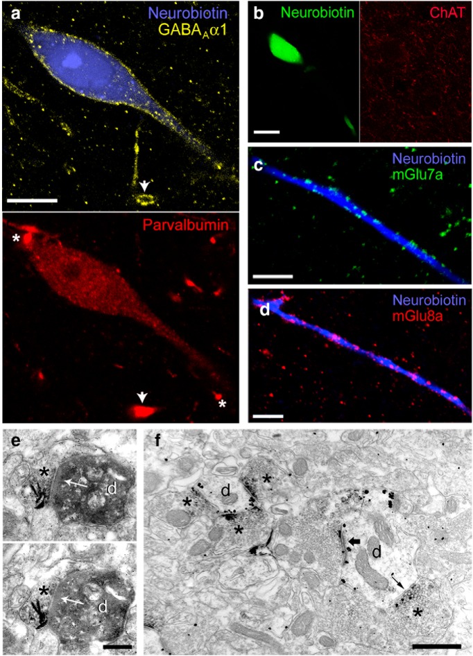
Figure 5.
Neurochemical profile of recorded L-ITCcs. a, Confocal micrographs showing strong expression of GABAA α1 (left) and moderate immunoreactivity for PV (right) in neuron tjx24a. Arrows denote nonfilled dendrites expressing comparatively higher levels of PV and GABAA α1; white asterisks indicate strongly PV-labeled nonfilled dendrites but devoid of GABAA α1 immunoreactivity. b, L-ITCcs are not cholinergic, as shown by the ChAT-negative biocytin-filled soma of tjx31a. c, Axon terminals containing mGlu7a receptors are apposed to the dendrites of L-ITCcs (tjx67a). d, Decoration of L-ITCc dendrites (tjx24a) by boutons containing mGlu8a receptors. e, Electron micrograph of a synapse (arrow) between a mGlu8a+ terminal (*) and a Neurobiotin-filled dendrite (d) of tjx24a (nickel-intensified DAB). f, Electron micrograph of a triple pre-embedding immunohistochemical reaction in which mGlu8a was visualized as a DAB-HRP reaction (dense precipitate *), whereas immunometal particles on postsynaptic dendritic (d) membranes revealed mGlu1α, and immunometal particles on presynaptic membranes revealed mGlu7a. Clear presynaptic labeling for mGlu7a can be observed in boutons forming both symmetric (i.e., potentially GABAergic, small arrow) and asymmetric synapses (i.e., potentially glutamatergic, large arrow) with mGlu1α+ dendrites. Note that mGlu7 and mGlu8 receptors are often expressed in the same terminals. Scale bars: a, 10 μm; b, 20 μm; c, d, 15 μm; e, 250 nm; f, 500 nm.

