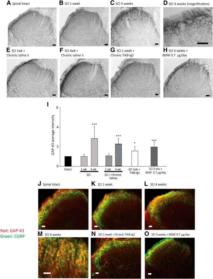
Figure 5.
Photomicrographs showing GAP-43 expression in the L5-L6 spinal cord segment of spinal-intact (A), 1 week (B), and 4 week (C) SCI animals; 1 week (E) and 4 week (F) SCI animals with an indwelling intrathecal catheter for saline delivery; 1 week SCI animals treated with chronic TrkB-Ig2 during the spinal shock (G); and 4 week SCI animals treated with chronic BDNF (0.7 μg/d for 28 d; H). Scale bars: A–C, E–H, 100 μm; D, 50 μm. GAP-43 was predominantly expressed in the superficial laminae of the dorsal horn, with a time-dependent increase after SCI (A–C), indicating the occurrence of fiber sprouting. GAP-43 immunoreaction was restricted to thin fibers running along the superficial dorsal horn (D). The expression of GAP-43 was not altered by chronic delivery of saline for 1 week (E) or 4 weeks (F). GAP-43 expression was significantly increased following administration of TrkB-Ig2 for 1 week (G) or BDNF (0.7 μg/d) for 4 weeks (F). I, Bar graph depicting the mean intensity of GAP-43 expression in the L5-L6 segment. The strongest intensity of GAP-43 was observed in 4 week SCI animals. The immunoreaction was also strong following BDNF sequestration TrkB-Ig2 compared with nontreated animals at the same time point. GAP-43 expression was unchanged in SCI animals that received chronic delivery of saline during 1 week or 4 weeks. BDNF administration induced a slight nonsignificant decrease of GAP-43 expression compared with nontreated 4 week SCI rats, but immunoreaction was still stronger than in spinal-intact animals (*p < 0.05, ***p < 0.001 vs spinal intact). J–O, GAP-43 expression colocalization with CGRP neuropeptide in the dorsal horn. Scale bar, 20 μm. In all experimental groups, there was a strict colocalization between GAP-43 and CGRP, which can be better observed in M.

