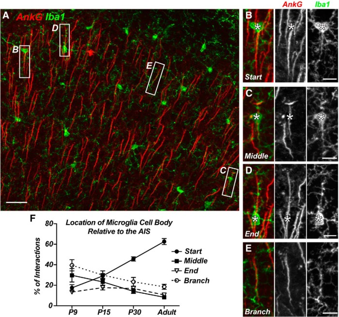
Figure 2.
AIS-associated microglial cell bodies are preferentially located at the start of the AIS. A, AnkG-labeled cortical AIS (red) and GFP+ microglia from a CX3CR1+/GFP mouse show multiple examples of AIS–microglia overlaps (white boxes). B–D, Microglial cell bodies located at the start (B), middle (C), and end (D) of the AIS. E, A “branched: microglial cell process associated with the AIS. F, Quantification of the location of the AIS-associated microglial cell body relative to the AIS at P9, P15, and P30 and in the adult. P9, P15, and P30 data are from M1 only; adult data include regions M1, S1BF, and V1. Scale bars: A, 25 μm; B–E, 8 μm.

