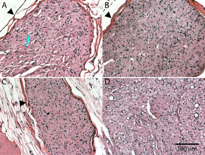
Figure 6.

Samples of (A) 8-week, (B) 16-week, (C) 24-week, and (D) 32-week posttreatment H&E-stained cross-sections of repeat-treatment sciatic nerves. Samples were collected at the indicated time intervals after the third cryotreatment. Arrow indicates degenerated axon. Arrowheads indicate epineurial structure of the nerve.
