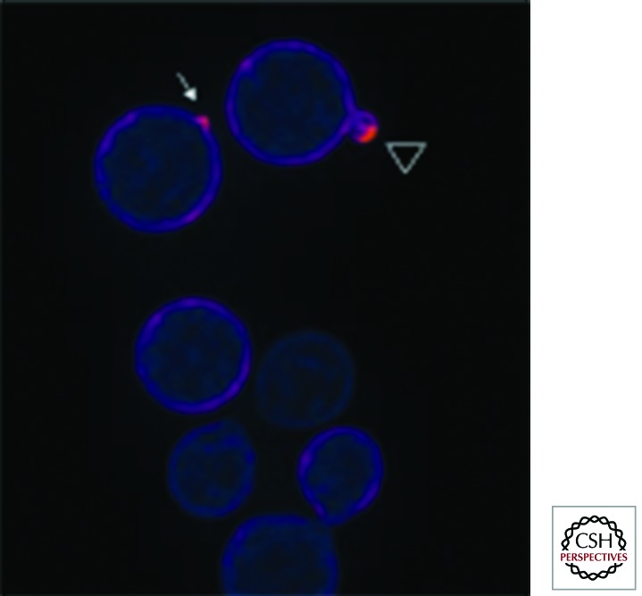
Figure 2.
C. neoformans Rac2 localization suggests a role in cell polarity. A Gfp–Rac2 fusion protein was expressed in C. neoformans and visualized using an Olympus (Center Valley, PA) IX70 microscope. After image acquisition and deconvolution of serial images in the Z-coordinate using Deltavision (GE Healthcare, Issaquah, WA) software, pseudocolored, merged images were produced using Fiji (Madison, WI) software. The most intense fluorescent signal, representing enrichment of Rac2 localization, is present at the site of incipient bud emergence (arrow) and distal edge of a small daughter cell (arrowhead). (Image provided by S. Esher and K. Selvig, Duke University.)

