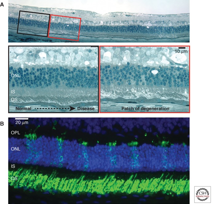
Figure 1.
Mosaic pattern of retinal degeneration in XLPRA2 female carriers. (A) Focal patches of photoreceptor degeneration surrounded by normally laminated retina in a 24-wk-old carrier. (B) Numerous patches of rod opsin mislocalization to the inner segments (IS) and outer nuclear layer (ONL), and features of rod neurite sprouting in the outer plexiform layer (OPL) in a 7-wk-old carrier. (Modified from data in Figure 1 from Beltran et al. 2009).

