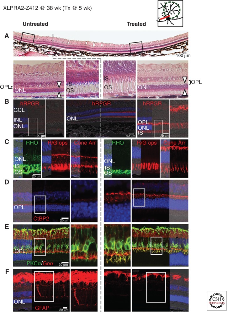
Figure 5.
Gene augmentation therapy with AAV2/5-hIRBP-hRPGRex1-ORF15 vector rescues photoreceptors when delivered at early-stage of XLPRA2 disease. Upper right diagram shows the area of subretinal injection performed at 5 weeks of age (green dotted line), and the site of the histological sections shown below at 38 weeks of age (red line) in XLPRA2 dog Z412. (A) H&E stained cryosection shows increased outer nuclear layer (ONL) thickness and inner (IS) and outer segment (OS) structure in the treated region. Dotted gray line shows the abrupt demarcation between the treated and untreated regions. (B) RPGR immunolabeling confirms expression of the transgene exclusively in the photoreceptors of the treated area and is seen throughout the cells with the exception of the outer segments. (C) Mislocalization of rod opsin (RHO, green) and red/green cone opsin (R/G, red) as well as cone morphology (cone arrestin, red) is corrected in the treated area. (D) Increased density of CtBP2/RIBEYE immunolabeled (red) synaptic ribbons in photoreceptor terminals and increased thickness of the outer plexiform layer (OPL) in the treated area. (E) Preserved dendritic arborization in rod bipolar cells immunolabeled with PKCα (green) and Goα (red) in the treated area. (F) Absence of Müller cell reactivity assessed by GFAP (red) immunolabeling in the treated area. (Modified from data in Figures S3 and 4 from Beltran et al. 2012).

