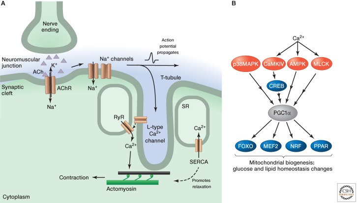
Figure 3.
Skeletal muscle contraction and changes with exercise. (A) Neurotransmitter (acetylcholine, ACh) released from nerve endings binds to receptors (AChRs) on the muscle surface. The ensuing depolarization causes sodium channels to open, which elicits an action potential that propagates along the cell. The action potential invades T-tubules and causes the L-type calcium channels to open, which in turn causes ryanodine receptors (RyRs) in the SR to open and release calcium, which stimulates contraction. Calcium is pumped back into the SR by (SR/ER calcium ATPase SERCA) pumps. The decreasing cytosolic calcium levels cause calcium to disassociate from troponin C and, consequently, tropomyosin reverts to a conformation that covers the myosin-binding sites. (B) Signaling in exercised skeletal muscle. Both calcium and calcium-independent signals stimulate the transcriptional coactivator PGC1α. This activates a number of transcription factors that regulate genes associated with mitochondrial biogenesis, glucose, and lipid homeostasis.

