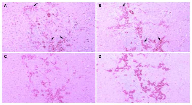
Figure 3.

Light microscopic distributions of immunohistochemically stained CAV-1 and -2 in PBC stage 3 liver tissue. A: CAV-1, ×100; B: CAV-2, ×100; C: double staining of CAV-1 and cytokeratin 7, ×100; D: double staining of CAV-2 and cytokeratin 7, ×100. Hematoxylin counterstain. Reaction products showing CAV-1 and -2 are localized abundantly on hepatic sinusoidal lining cells and proliferative bile ductules. Regenerating bile ductules at the interface of portal tracts and necrotic areas are immunostained intensely for CAV-1 and -2, showing significant increase compared to control liver. Arrow denotes proliferative bile ductule.
