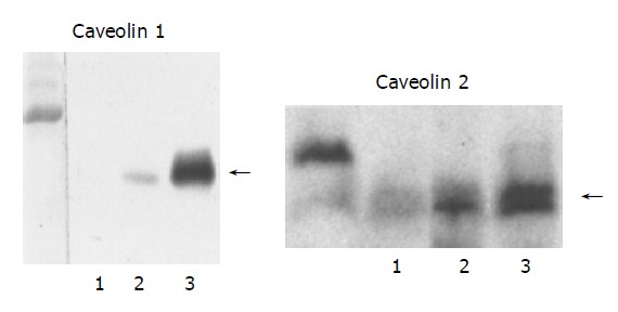
Figure 4.

Western blot analysis of the expression of CAV-1 and -2 proteins in human control and PBC liver tissues. Lane 1: normal liver; lane 2: PBC stage 1 liver; lane 3; PBC stage 3 liver. CAV-1 and -2 are found in abundance in PBC stage 3 specimens, less intensely in PBC stage 1, and is undetectable in normal liver tissue. Samples containing 50 μg of membrane protein were subjected to electrophoresis on SDS/PAGE gel (CAV-1 and -2: 4-20%) and analyzed by blotting.
