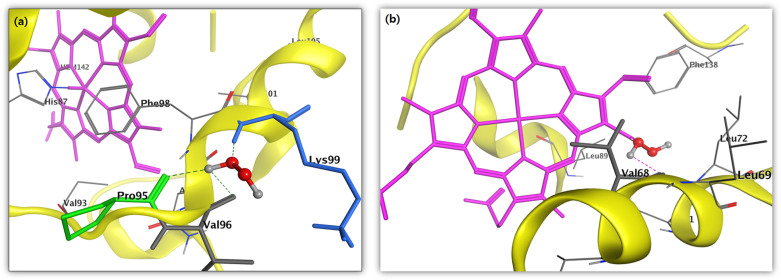
Figure 4. In silico molecular docking showing the binding site residues within a selection radius of 4 Å from bound H2O2 revealing the hydrophobic pocket of active conformation.
Binding sites of human (a) Hb (PDB:1A3N) and (b) Mb (PDB:3RGK) protein receptor. The docking studies were carried out using SYBYL-X 2.0.

