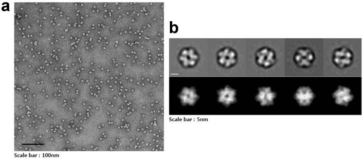
Figure 2. Dodecameric structure of CpsUbiX.
(a) A typical micrograph depicting the CpsUbiX dodecamer. (b) Representative two-dimensional (2D) class averages of the CpsUbiX dodecamer and corresponding forward projection images. Negative-staining electron micrographs of the purified recombinant CpsUbiX showed ball-shaped structures approximately 10 nm in length. The scale bar is 5 nm.

