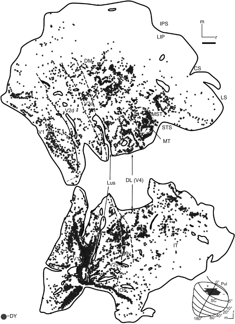
Figure 3.
DY-labeled cells in case 1.
Notes: Black circles indicate the distribution of DY-labeled cells through the right visual cortex of case 1. Areal boundaries were determined from adjacent sections stained for cytochrome oxidase and myelin. Thin black lines are borders determined from anatomical sections. Dashed lines indicate borders that were determined from both anatomical sections and measurements. The finest dashed line is the horizontal meridian. Thick black lines denote tears and the edges of the block. In cortex, right is rostral, up is medial. Scale bar is 5 mm.
Abbreviations: CS, central sulcus; DL, dorsolateral area; DM, dorsomedial area; DY, diamidino yellow; IOS, inferior occipital sulcus; IPS, intraparietal sulcus; IT, inferior temporal cortex; LIP, lateral intraparietal area; LS, lateral sulcus; LuS, lunate sulcus; MST, medial superior temporal area; MT, middle temporal area; Pul, pulvinar; SC, superior colliculus; STS, superior temporal sulcus; d, dorsal; v, ventral.

