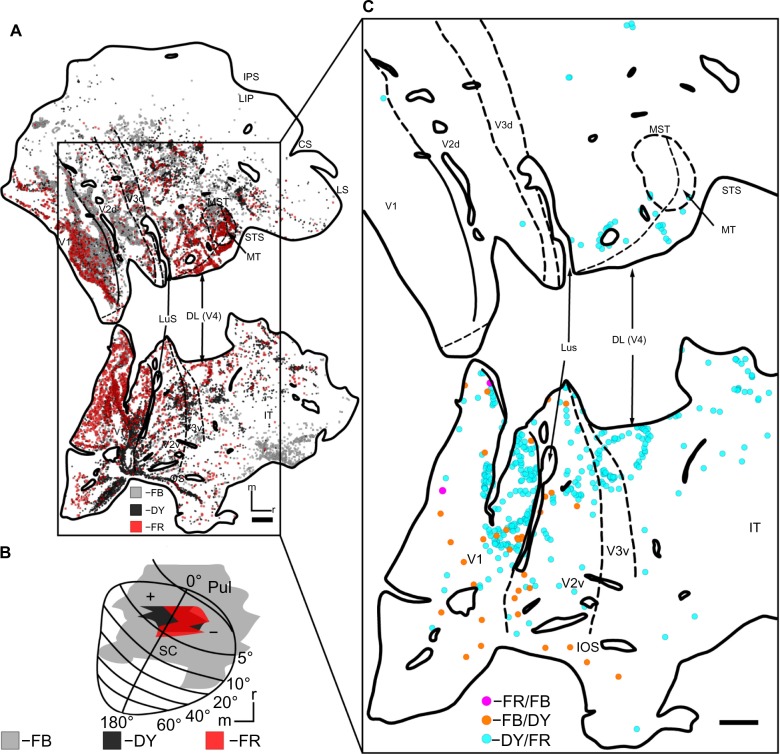
Figure 6.
Double-labeled cells in case 1.
Notes: (A) Distribution of single-labeled cells through the right visual cortex of case 1. In cortex, right is rostral, up is medial. (B) Top-down view of the reconstructed case 1 injection sites in the SC. Left is medial, up is rostral. (C) Distribution of double-labeled cells through the right visual cortex of case 1. Scale bar is 5 mm.
Abbreviations: CS, central sulcus; DL, dorsolateral area; DY, diamidino yellow; FB, fast blue; FR, Fluororuby; IOS, inferior occipital sulcus; IPS, intraparietal sulcus; IT, inferior temporal cortex; LIP, lateral intraparietal area; LS, lateral sulcus; LuS, lunate sulcus; MST, medial superior temporal area; MT, middle temporal area; Pul, pulvinar; SC, superior colliculus; STS, superior temporal sulcus; d, dorsal; v, ventral.

