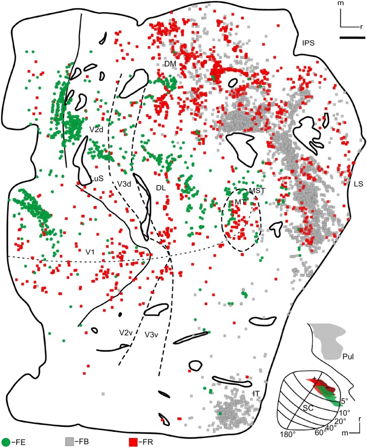
Figure 7.
The distribution of cells through the right visual cortex of case 2.
Note: In cortex, right is rostral, up is medial. Scale bar is 5 mm.
Abbreviations: DL, dorsolateral area; DM, dorsomedial area; FB, fast blue; FE, fluoroemerald; FR, Fluororuby; IPS, intraparietal sulcus; IT, inferior temporal cortex; LS, lateral sulcus; LuS, lunate sulcus; MST, medial superior temporal area; MT, middle temporal area; Pul, pulvinar; SC, superior colliculus; d, dorsal; v, ventral.

