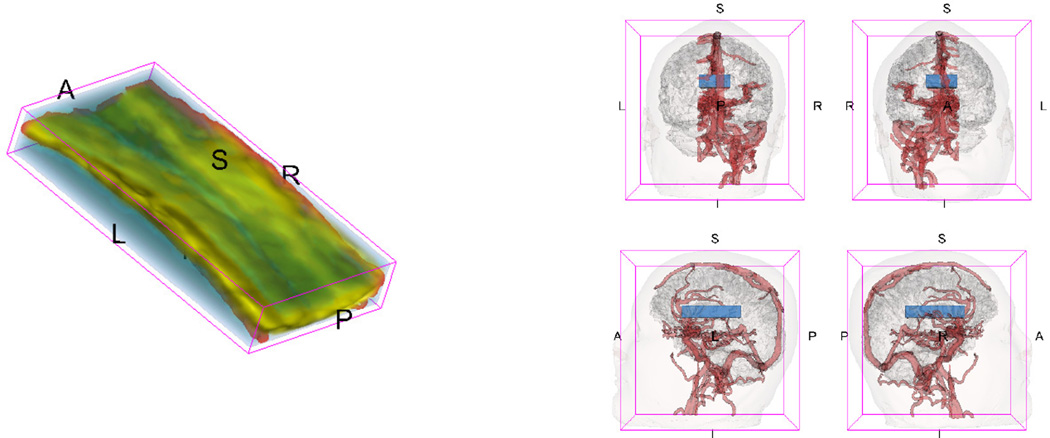
Figure 1.
The 3D-rendering of the region of interest (left), a blue block containing corpus callosum, and the template brain (right). Views: R=Right, L=Left, S=Superior, I=Interior, A=Anterior, P=Posterior. For the purposes of orientation, major venous structures are displayed in red in the right half of the template brain. The 3D-renderings are obtained using 3D-Slicer (2011) and 3D reconstructions of the anatomy from Pujol (2010).

