Figure 1.

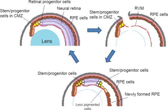
A schema for retinal regeneration in X. tropicalis: stem/progenitor cells in the ciliary marginal zone regenerate the whole retina.
(A) Normal eye. During the growth of the eye, stem/progenitor cells in the ciliary marginal zone (CMZ) produce retinal progenitor cells, which are incorporated into the retina at the periphery. (B) An eye immediately after retinal removal. An incision is made at the dorsal periphery of the eye, and the lens fibers are removed, leaving the retinal vascular membrane (RVM) in the posterior chamber. Some of the retinal pigmented epithelial (RPE) cells begin to be detached and migrate to the RVM, where they form a new RPE layer (as seen in Figure C). (C) At the later stage of regeneration (days 15–20), the regenerating retina extends further to the posterior. At the posterior edge of the regenerating retina, a proliferating zone (progenitor zone) is observed (consisting of actively proliferating cells). At the peripheral end of the regenerating retina (closer to the iris) there still remain progenitor cells that are intensely labeled for BrdU. The regenerating retina is sandwiched by progenitor cell zones at both ends. In X. tropicalis, RPE cells in the newly formed epithelium do not appear to transdifferentiate into neuronal cells, although some cells at the boundary to the regenerating retina become less pigmented. These cells are gradually lost as the regenerating retina extends more posteriorly. Contrary to X. tropicalis, the RPE cells on the RVM transdifferentiate into the retina in X. laevis. The schema is modified from the original figure (Miyake and Araki, 2014).
