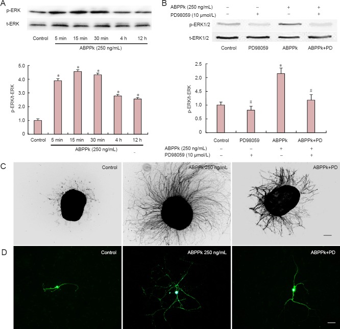
Figure 3.
ABPPk-induced activation of phosphorylation of ERK1/2 in cultured DRG neurons.
Bar graphs illustrating the ratio of p-ERK/t-ERK in DRG neurons treated with 250 ng/mL ABPPk for different times (A) or treated with 10 μmol/L PD98059 (a specific inhibitor of Erk1/2) for 30 minutes and then treated with 250 ng/mL ABPPk for different times (B). The data are presented as the mean ± SEM of three separate experiments (each in triplicate). *P < 0.05, vs. control group; #P < 0.05, vs. ABPPk treatment alone (one-way analysis of variance plus Scheffé post hoc test). Also shown (inset) are the representative western blot images. Micrographs obtained after DRG explants (C) or DRG neurons (D), which had been cultured for 72 hours in plain medium (control), or in the medium added with 250 ng/mL ABPPk in the absence or presence of 10 μmol/L PD98059, and were immunostained with anti-GAP43 (C) and anti-β-tubulin III (D). Scale bars: 20 (A) and 50 (B) μm. ABPP: Achyranthes bidentata polypeptides; DRG: dorsal root ganglion; GAP43: growth-associated protein 43.

