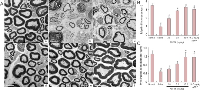
Figure 4.

Nerve regeneration in mice after sciatic nerve crush injury.
Transmission electron micrographs obtained at 21 days after surgery, showing the regenerated nerves (A) at the contralateral, normal side (a) and crushed side of animals receiving injection of saline (b), 2.5 mg/kg (c), 5.0 mg/kg (d), 10.0 mg/kg (e) of ABPPk, and 16.0 mg/kg of ABPP (f). Scale bars: 5 μm. Bar graphs comparing the myelin thickness (B) and the myelinated axon diameter (C) from the normal side and at the crushed side of mice receiving different treatments as indicated. All data are presented as the mean ± SD (n = 10). *P < 0.05, vs. saline treatment; #P < 0.05, vs. normal side (one-way analysis of variance plus post hoc Scheffé test); ABPP: Achyranthes bidentata polypeptides.
