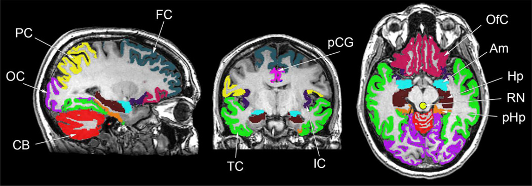
Figure 2.
Regions used for analysis of 18F-mefway PET data. Regions defined by the template-based FreeSurfer algorithm included the amygdala (Am), hippocampus (Hp), parahippocampal gyrus (pHp), insular cortex (IC), anterior cingulate gyrus (aCG; not shown), posterior cingulate gyrus (pCG), parietal cortex (PC) orbitofrontal cortex (OfC), temporal cortex (TC), occipital cortex (OC), frontal cortex (FC). The hand drawn raphe nuclei (RN) is also shown.

