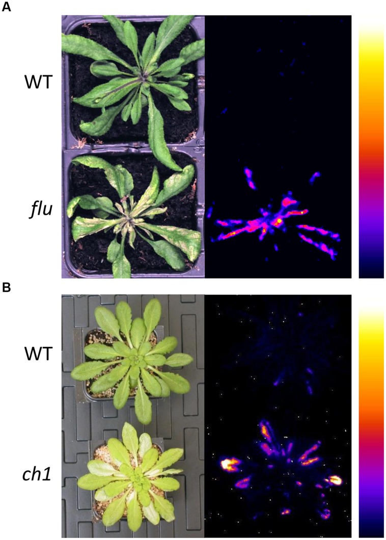
FIGURE 1.
Photosensitivity of the 1O2-overproducing Arabidopsis mutants, flu and ch1. (A) Wild-type (WT) and flu mutant plants after an 8 h dark/light transition, showing leaf damage (2 days after the dark-to-light shift, left) and lipid peroxidation as measured by autoluminescence imaging (2 h after the dark-to-light shift, right). (B) WT and ch1 mutant plants after high light stress (1000 μmol photons m-2 s-1 for 2 days), showing leaf bleaching (left) and lipid peroxidative damage (right). No luminescence and no leaf damages were detected in WT and the mutant plants before the light treatments. The color palette on the right indicates the intensity of the luminescence signal from low (dark blue) to high (white). The signal intensity is indicative of the amount of lipid peroxides present in the sample (Birtic et al., 2011). Adapted from Ramel et al. (2013a) (Copyright American Society of Plant Biologists, http://www.plantcell.org) and completed.

