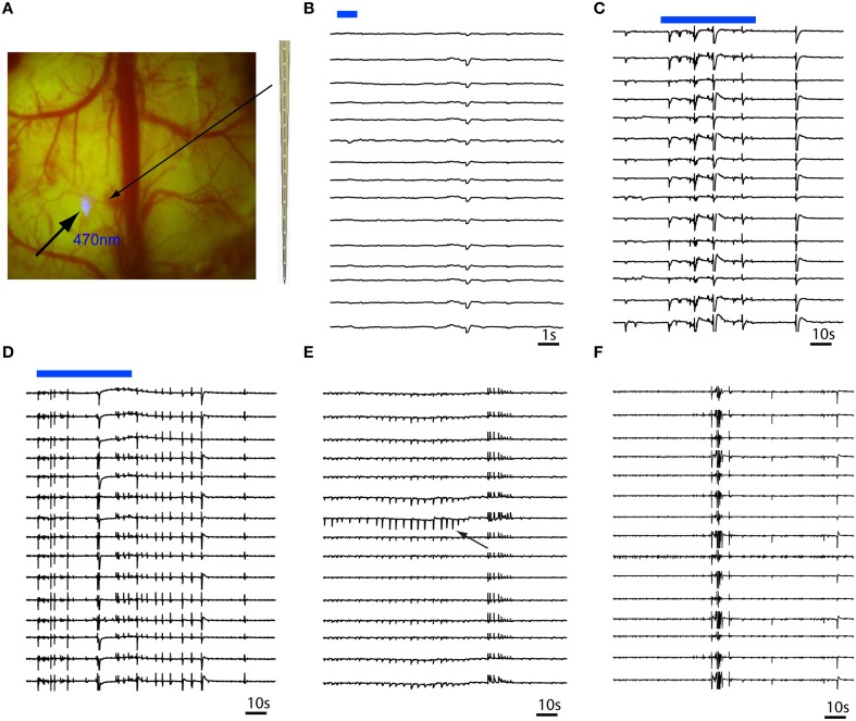
Figure 3.
Multi-contact laminar depth array recording of in vivo RuBi-4-AP epileptic events. The depth electrode is placed 0.2 mm away from the site of focal illumination. (A) Focal photostimulation 0.2 mm away from the laminar depth electrode placed tangentially into the cortex. (B) A 1 s pulsed photostimulation (blue line) results in a short duration event that occurred simultaneously in all layers. (C,D) Longer duration illumination (60 s) results in immediate onset of polyspikes and short duration events involving all layers simultaneously. (E) Occasionally, layer specific events (black arrow) occur that quickly spread to all other layers. (F) However, the majority of events involve all layers simultaneously. The blue bars in (B,C) show the timing of blue photostimulation.

