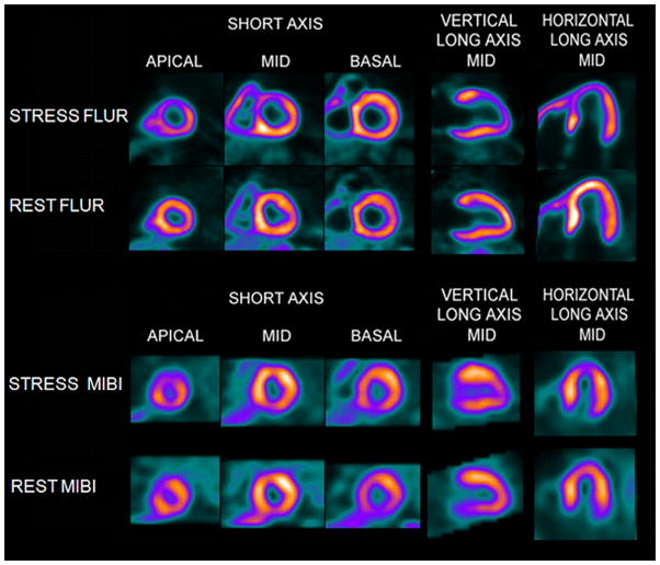
Figure 7.
Flurpiridaz F 18 (FLUR) PET(top) and Tc-99m sestamibi (MIBI) SPECT(bottom) images from an 82 year old man with shortness of breath and an occluded native proximal left anterior descending (LAD) coronary artery and an occluded left internal mammary graft to the LAD and no other significant native coronary artery disease. The FLUR images show a severe reversible perfusion defect throughout the territory of the occluded proximal LAD, while the MIBI images show only a moderate perfusion defect in the distal LAD territory (apical slices).

