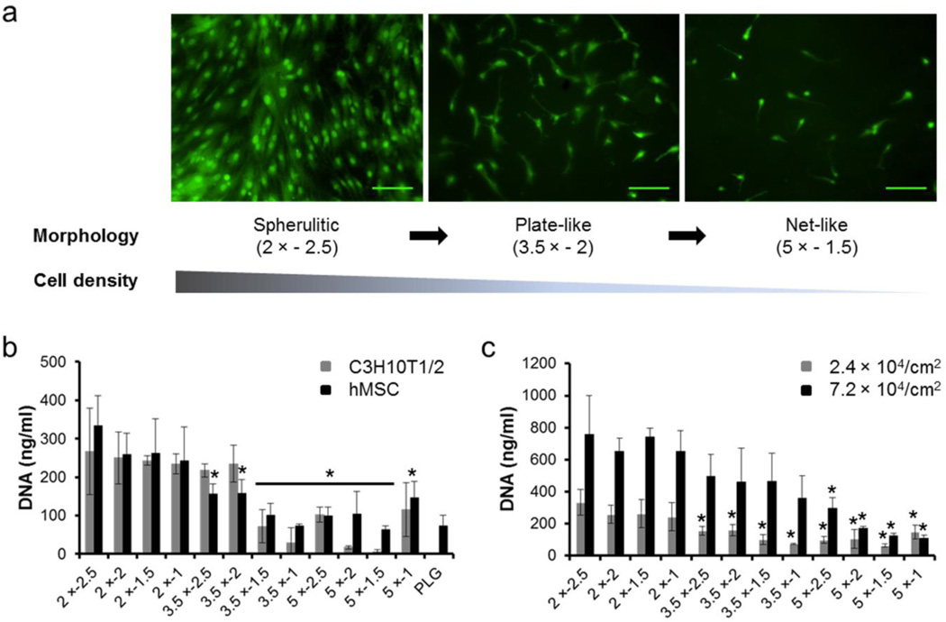
Fig. 5.
Multipotent stem cell expansion on mineral coatings. (a) Fluorescence images of hMSC after a 4 day incubation on 2 × − 2.5 (left), 3.5 × − 2 (middle) and 5 × − 1.5 (right) mineral coatings. Scale bar = 100 µm. (b) hMSC and C3H10T1/2 cells expansion on mineral coatings and PLGA surface. Total DNA amount (a measure of cell number) of cells after 8 day incubations. (c) Seeding density effect on hMSC expansion after 8 day incubations. * indicate significant difference when compared with 2 × − 2.5 conditions respectively.

