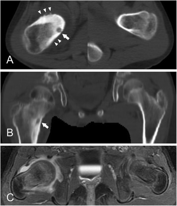
Figure 1.

A 9.7-year-old girl with a juxta-articular osteoid osteoma of the proximal femur with involvement of the hip joint. (A) Axial CT image of the right femoral neck shows a prominent bony hypertrophy (arrowheads) around the nidus (arrow). (B) Coronal CT image demonstrates a nidus just above the lesser trochanter of the femur (arrow). Note the widening of the femoral neck and hypertrophy of the femoral head compared with the contralateral normal side. (C) Axial contrast-enhanced T1-weighted MR image with fat suppression shows synovial hypertrophy with effusion. The head-neck offset is not clear.
