
FIGURE 1.
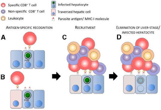
Model of the elimination of malaria liver stage by a CD8+ T cell-dependent inflammatory cell cluster. (A) Parasite antigens cross the parasitophorous vacuole membrane (PVM), are processed and presented on the surface of the infected hepatocyte by MHC I molecules, and/or (B) are released in the cytoplasm and presented on the surface of hepatic cells (hepatocytes, Kupffer cells, endothelial cells—figuratively represented by the pink cells) traversed by sporozoites. (A,B) Parasite-specific, activated CD8+ T cell (red cell) recognizes its cognate antigen and (C) recruits other specific and non-specific (purple cells) CD8+ T cells, and presumably other leukocytes (orange cells) to the site of infection. (D) These immune cells cluster around the infected hepatocyte, leading to the elimination of the malaria liver stage and presumably of the infected hepatocyte. For clarity and due to the lack of information about its position during cluster formation, the sinusoidal barrier is not represented in the model.