
Figure 1. Proteomic profiling of cancer cells.
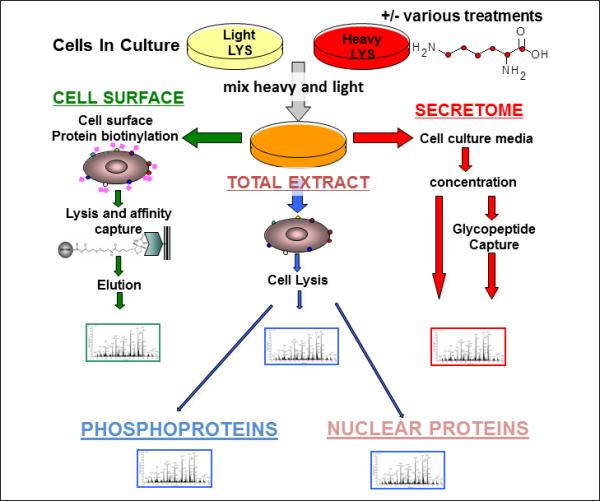
Freshly isolated tumor cells or cancer cell lines to be compared are cultured in the presence of isotopically labeled amino acids. Media, biotinylated cell surface proteins and whole lysates are isolated separately and analyzed. Cell lysates may be further fractionated for separate analysis of sub-compartments (e.g. nuclear proteins), or protein subgroups (e.g. phosphoproteins).