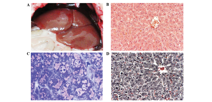
Figure 5.
Images of a GIII rat. (A) Gross morphology of the liver of a GIII rat with a normal reddish-brown color. (B) Hematoxylin and eosin-stained section of the liver. The image is devoid of fibers and lipid droplets (magnification, ×200). (C) Toluidine blue-stained section of the liver. The image is devoid of fibers and lipid droplets (magnification, ×1,000). (D) Masson’s trichrome-stained section of the liver with no collagen fibers in the extracellular matrix. The image also lacks lipid droplets (magnification, ×400). GIII, carbon tetrachloride plus ethanol plus green tea extract group.

