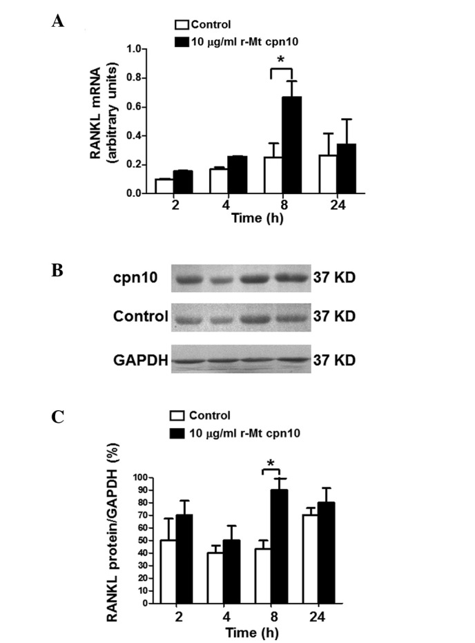
Figure 4.

Levels of RANKL in third-generation osteoblasts following treatment with r-Mt cpn10 (10 μg/ml) for 2, 4, 8 and 24 h. (A) Levels of RANKL mRNA following stimulation with control or r-Mt cpn10 (10 μg/ml) for 2, 4, 8 and 24 h. Total RNA was isolated from the harvested cells, transcribed to cDNA and quantified by reverse transcription-quantitative polymerase chain reaction analysis. The amount of RANKL mRNA was compared with the amount of the reference gene (GAPDH) mRNA. Values are expressed as the mean ± intra-assay variation of duplicate analyses. (B) Western blot analysis of RANKL protein expression following treatment with the control or r-Mt cpn10 (10 μg/ml) for 2, 4, 8 and 24 h. (C) Quantification of the level of RANKL protein following treatment with the control or r-Mt cpn10 (10 μg/ml) for 2, 4, 8 and 24 h. Results are expressed as the mean ± standard devation. *P<0.05. RANKL, receptor activator of nuclear factor-κB ligand; r-Mt, recombinant Mycobacterium tuberculosis; cpn10, 10-kDa co-chaperonin.
