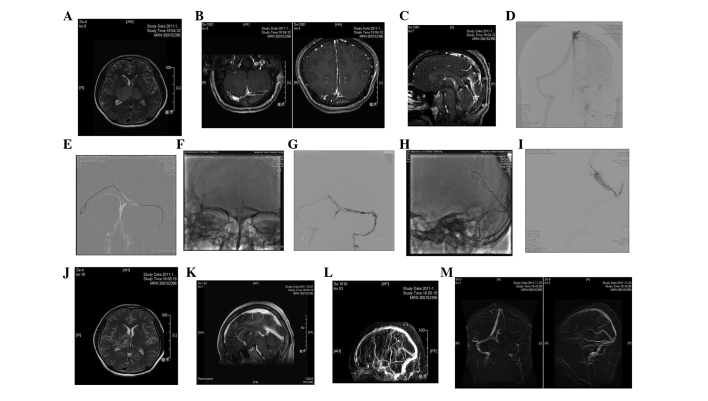
Figure 2.
Catheter-directed thrombolysis in the straight sinus. (A) Head MRI T2 weighted image showed that the bilateral thalami were infarcted and the right side was more severe; (B) thrombus in the straight sinus as shown by enhanced MRI; (C) thrombus in the left transverse sinus and superior sagittal sinus as shown by enhanced MRI; (D) DSA showed that the left transverse sinus and straight sinus did not develop; (E) guidewire entered the transverse sinus during surgery; (F) sacculus expanded during surgery; (G) left transverse sinus was open during surgery; (H) microcatheter was placed in the straight sinus; (I) microcatheter angiography revealed that the straight sinus developed following thrombolysis; (J) 2 weeks after surgery, review of the T2 weighted image showed that disease extent of the thalamus had reduced; (K) enhanced imaging showed that the superior sagittal sinus and straight sinus were open; (L) MRV showed that the superior sagittal sinus and straight sinus were open; (M) 6 weeks after surgery, MRV showed that the straight sinus was open but the left transverse sinus was closed. CT, computed tomography; MRI, magnetic resonance imaging; DSA, digital subtraction angiography; MRV, magnetic resonance venography.

