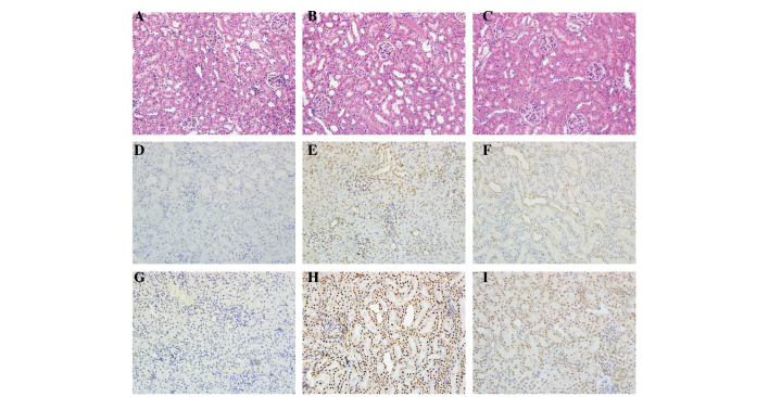
Figure 2.
Histological features were evaluated by H&E and TUNEL staining and immunohistochemistry was performed to evaluate the expression of cleaved caspase-3. Representative kidney sections stained with (A-C) H&E, (D-F) TUNEL and (G-I) cleaved caspase-3 (brown nuclear staining) in kidneys at the end of the 24 h reperfusion period. Sections from (A,D,G) a sham-operated rat, (B,E,H) a rat subjected to I/R and (C,F,I) a rat subjected to picroside II treatment. All images of H&E, TUNEL and immunohistochemical staining, original magnification ×200. H&E, hematoxylin and eosin; TUNEL, terminal deoxynucleotidyl transferase-mediated deoxyuridine triphosphate-biotin nick end labeling; I/R, ischemia and reperfusion.

