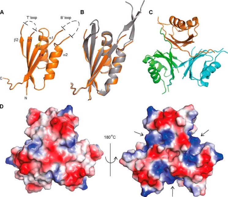
FIGURE 2.
Crystal structure of apo-PstASA. A, schematic representation of the crystal structures of a PstASA monomer with α-helices and β-strands numbered, and unstructured T′- and B′-loops schematically indicated; B, overlay between PstASA (orange) and the P. pentosacues ATCC 25745 protein PEPE_1480 (PDB code 3M05) (gray); and C, PstASA trimer with the individual monomers shown in different colors. D, electrostatic surface potential representation (blue, positive potential; red, negative potential) of the PstASA trimer shown on the left in the same orientation as in schematic representation in C and on the right rotated by +180° around the y axis.

