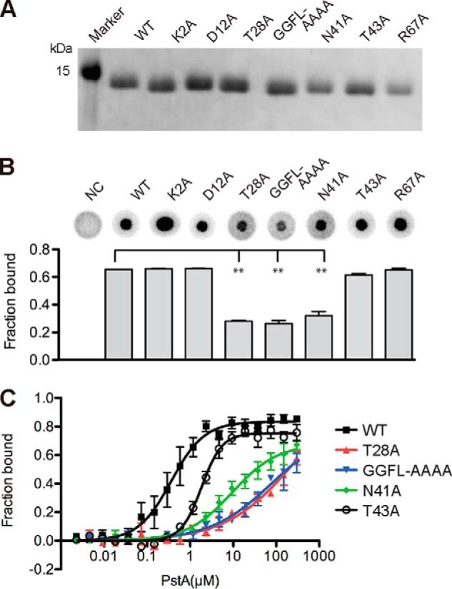
FIGURE 8.

Identification of PstA variants with c-di-AMP-binding defects. A, Coomassie-stained gel with purified His-PstASA protein (WT) and different protein variants containing the amino acid substitutions as indicated above each lane. B, c-di-AMP binding assays. DRaCALAs were performed with 32P-labeled c-di-AMP and purified His-PstASA protein (WT) or variants with the indicated amino acid substitution. The proteins were used at a concentration of 10 μm, and as negative control (NC) no protein was added to the binding assays. Representative DRaCALA spots are shown, and the fraction bound was calculated as described previously, and the average values and standard deviation of two independent experiments were plotted. C, binding curve and Kd determination for c-di-AMP and purified His-PstASA or protein variants with the indicated amino acid substitutions. Kd values were determined from the curve as described previously (41).
