
FIGURE 9.
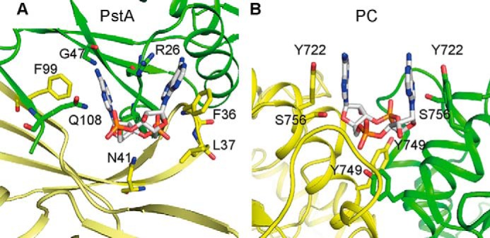
Comparison of S. aureus PstA and L. monocytogenes PC c-di-AMP-binding sites. A and B, schematic representations of c-di-AMP-binding site in S. aureus PstASA (A) and L. monocytogenes PC (PDB 4QSH) (11) (B) with different monomers shown in green and yellow, respectively. Amino acid residues forming key contact with the ligand are labeled and shown as sticks.