FIGURE 1.
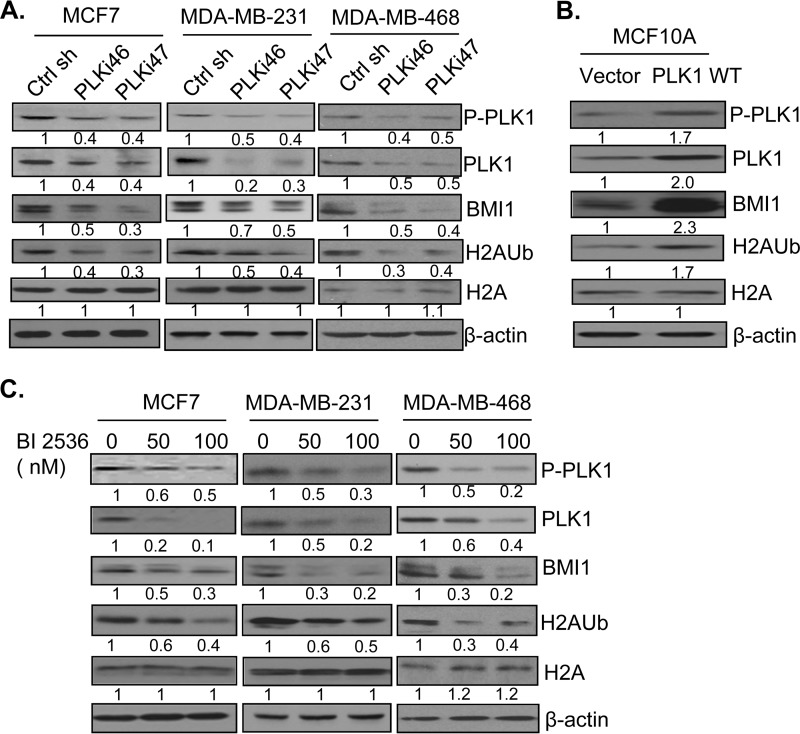
PLK1 regulates expression of the PcG protein BMI1 and PRC1 activity. A, MCF7, MDA-MB-231, and MDA-MB-468 cells stably expressing a control and two PLK1-specific shRNAs (PLKi46 and PLKi47) were generated by infection with respective retroviral vectors and selection in puromycin (1 μg/ml). The expression of PLK1, phospho-PLK1 (P-PLK1), BMI1, total H2A, H2AK119Ub, and β-actin was determined by Western blot analysis. B, MCF10A cells overexpressing PLK1 were generated by infecting cells with PLK1 expressing retrovirus, and selecting cells in G418 for 7 days. After selection, control (vector) and PLK1 WT-expressing cells were analyzed for PLK1, P-PLK1, BMI1, total H2A, H2AK119Ub, and β-actin by Western blot analysis. C, indicated sets of cells were treated with 0 (DMSO), 50, and 100 nm of PLK1 inhibitor BI 2536 for 24 h. The total cell lysates were prepared and expression of PLK1, P-PLK1, BMI1, total H2A, H2AK119Ub, and β-actin was detected by Western blot analysis. The proteins were quantified by densitometry using ImageJ software and the fold induction of each protein normalized to β-actin was determined. The fold induction of relevant protein is shown below the immunoblot (IB) of each protein.

