
FIGURE 2.
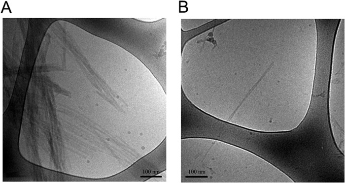
Formation of α-syn fibrils. 5 μl of α-syn fibrils in 10 mm MES, pH 5.5, with 140 mm NaCl, unless otherwise stated) was added to the glow-discharged grid for cryo-TEM imaging. A, examples of typical α-syn fibrils formed at pH 5.5. B, example of a typical α-syn fibril formed at pH 7.5.