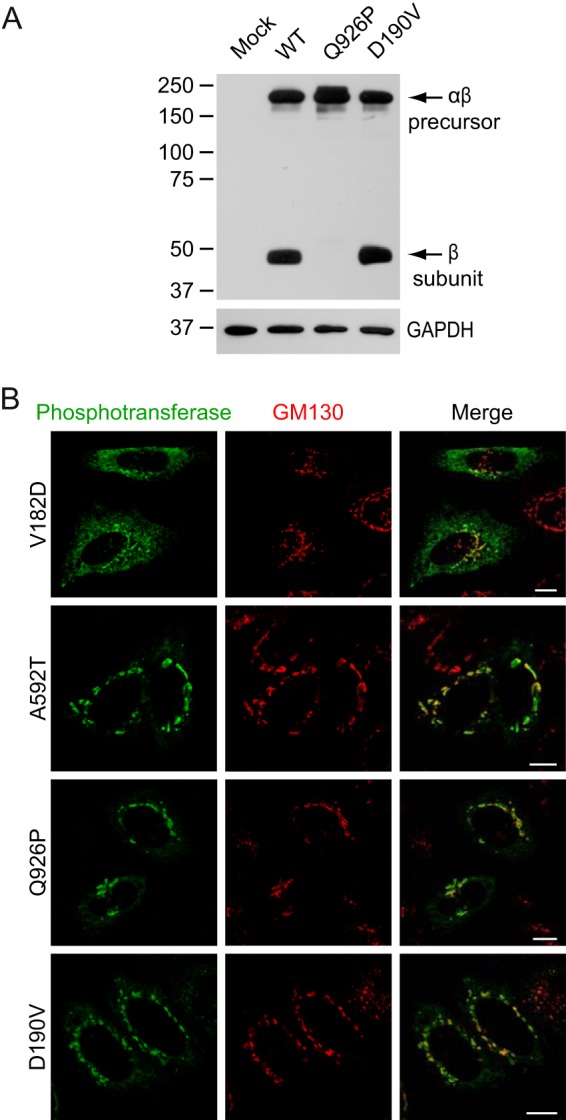
FIGURE 7.

Consequences of mutations in the spacer regions of αβ phosphotransferase. A, Western blots (anti-V5) of HEK293 cell lysates ∼48 h after transfection with WT or mutant phosphotransferase αβ subunits with a C-terminal V5 tag. B, confocal immunofluorescence microscopy shows the subcellular localization of several spacer mutants in HeLa cells. Note that the V182D mutant shows partial ER localization (anti-α in green) and overlaps with the cis-Golgi marker GM130 (red). A592T αβ phosphotransferase has a predominant Golgi localization. Scale bars, 10 μm.
