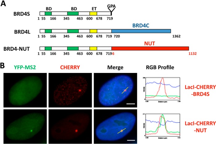
FIGURE 1.
NUT protein activates in situ gene transcription. A, schematics of the BRD4 isoforms and BRD4-NUT oncogenic fusion. The t(15;19) translocation breakpoint bisects BRD4L at amino acid 719. The N-terminal component contains the double bromodomains (BD), a potential kinase domain, an extraterminal (ET) protein-protein interaction domain, and a serine-rich domain. The C-terminal of BRD4-NUT incorporates almost the entire NUT sequence. The BRD4C region spanning the BRD4 C-terminal amino acids 720–1362 is specifically present in BRD4L but not in BRD4-NUT. B, U2OS 2-6-3 YFP-MS2 stable cells were transfected with LacI-CHERRY-BRD4S or LacI-CHERRY-NUT. 24 h post-transfection, cells were fixed and counterstained with DAPI. LacI-CHERRY fusions allow the LacO locus to be visualized in red, whereas YFP-MS2 marks the sites of active transcription. IF, immunofluorescent staining with indicated antibodies (p300, CBP, AcH4, or BRD4). The RGB intensity profiles show the intensity curve of red, green, and blue signals along the highlighted yellow bars in the Merge panel. Scale bars = 5 μm.

