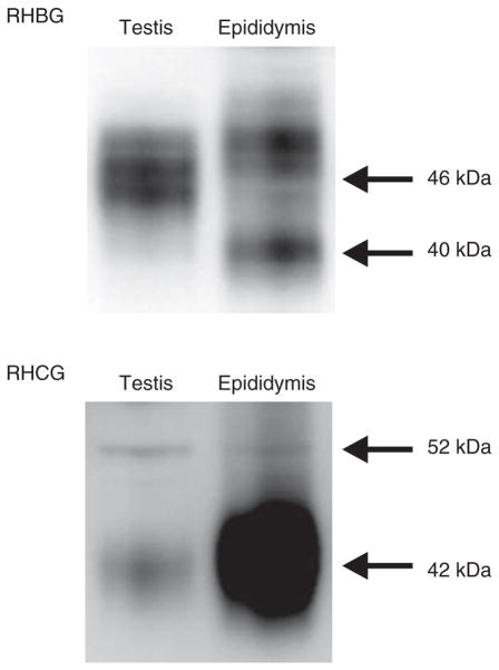
Figure 2.
Immunoblot analysis of RHCG and RHBG in the male reproductive tract. (Upper panel) Immunoblot analysis of RHBG protein expression. RHBG is present in both testis and epididymis.
The predominant expression of RHBG is at ~46 kDa in the testis and 40 and 49 kDa in the epididymis, consistent with tissue-specific variations in glycosylation. (Lower panel) Immunoblot analysis of RHCG protein expression. The predominant expression of RHCG is at 42 and 52 kDa in the testis and at 42 kDa in the epididymis. These variations are consistent with tissue-specific variations in RHCG glycosylation.

